WLC 8TH GRADE SCIENCE CLASS'S AWESOME WEBSITE
CHAPTER 14 TEST

Home

PERCH PRELAB PRINT THIS | CONTACT MR.FOX | PERCH LAB PRINT THIS | PERCH REVIEW | CAT MUSCLES | CHORDATES | FISH SCALES | HOMEWORK | CLASSWORK | BIOMES | BIOME PROJECT | CHAPTER 14 TEST | CHAPTER 14 TEST

  Starfish dissection (aboral surface)

Lab_8b-16a

  1. Arm (ray)

  2. Gonad

  3. Stomach

  4. Pyloric cecum

FredsLine

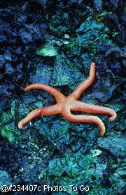
This image shows a preserved starfish with a portion of the aboral surface of one arm (ray) removed.  The dissection mount shows the central stomach and large pyloric caecum that occupies most of the space within each arm as well as the smaller gonads.

FredsLine


Lab_8b-17a

  1. Ambulacral groove

  2. Mouth

FredsLine

This image shows the oral surface of a preserved starfish.  Note the central mouth and ambulacral groove that runs down the center of the oral surface of each arm.  Extending from each ambulacral groove are numerous tube feet that are used for locomotion, attaching to the substrate and feeding. 


Lab_8b-18a

  1. Central disk

  2. Arm

  3. Madreporite

  4. Ambulacral ossicles

  5. Endoskeletal plates

  6. Ampullae

  7. Coelom

  8. Anus

  9. Pyloric stomach

10. Cardiac stomach

11. Digestive gland

12. Gonad

13. Stone canal

FredsLine

This image shows a partially dissected view of the aboral surface of a plastic model of a starfish.  The image contains the principal internal organs and elements of the water-vascular system,  including the madreporite (the entrance to the system), the stone canal and the ampullae, which deliver fluid under pressure to extend the tube feet so that they can make contact with the substrate or a food item. 

Enter subhead content here

Sea Star PICTURES

Dissecting starfish is fun but often frustrating, especially when working with preserved specimens.  The following pictures are of a typical, CHEAP, preserved starfish: the kind my students usually end up dissecting.


Here's a dorsal view showing the madreporite (sieve plate).


This is a closer view of the madreporite and the anal area.


The pedicellariae are very tiny clawlike structures that help keep this echinoderm's skin clean.


Here's a ventral view showing the mouth area and the ambulacral groove where the tube feet are found.


This is a closeup of the mouth area showing the ends of the tube feet.


Here's what the end of a cut-off arm looks like.  It's pretty difficult to distinguish the structures in a preserved specimen like this one.


With the top removed, you can see the stomach and digestive glands.


Remove the stomach and glands to see the ring and radial canals of the water vascular system.


A closeup of the opened arm shows the ampullae that are the top ends of the tube foot structures.


Enter content here

Enter content here

Enter content here

Enter supporting content here